
Right shoulder anatomy
Full 360 exploration mode
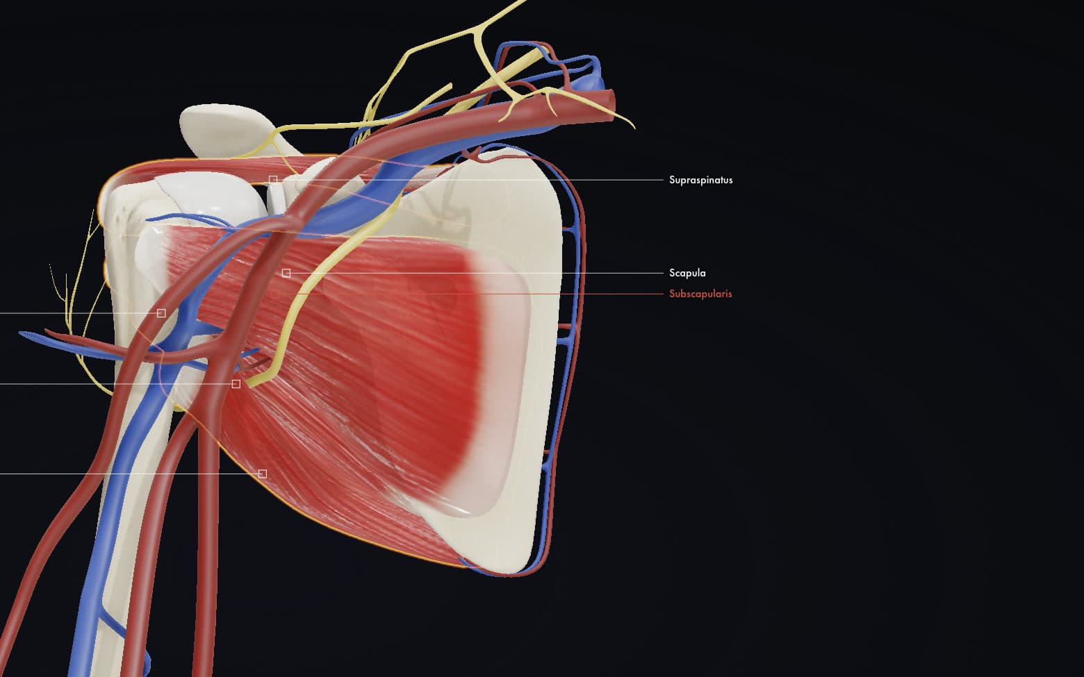
A real-time 3D right shoulder you turn, fade and take apart yourself: bones, cartilage, the four rotator cuff muscles, and the vessels and nerves around them.
A CDI Interactive experience by CDI StudioUpdated
It runs in its own tab so it gets the whole screen, the sound and the full frame rate.
Overview
Right Shoulder: Anatomy is the first module in the CDI Interactives series. It is a real-time 3D shoulder you explore yourself, in a browser, with nothing to install and no account to create. A video decides what you see and how long you see it for. This does not. Turn the joint to any angle, fade a layer down to see what sits behind it, switch individual structures on and off, and stay on one part for as long as you need. It runs on a phone, a laptop or a lecture room display, from a single link.
The Science
The shoulder buys its range of movement by giving up bony stability. The humeral head sits against the glenoid, a socket far shallower than the head it holds, and the joint stays together because soft tissue holds it there rather than because the bone traps it. That trade is the reason the shoulder moves further than any other joint in the body and the reason it is the one that most often gives way.
Four muscles do the holding. Subscapularis covers the front of the scapula and turns the arm inwards; supraspinatus runs over the top and starts the arm away from the body; infraspinatus and teres minor sit behind and turn it outwards. Together they pull the head into the socket through the whole arc of movement. Teres major lies alongside them and is often mistaken for one of the four; the model carries it so the difference can be seen rather than described.
Around and through all of it run the vessels and nerves that make the region unforgiving to operate in. Switching the muscle layer off and the nerve layer on shows why: the nerve supply wraps the bone closely enough that the anatomy, not the textbook, is what explains the risk.
How to Interact
Pick a system. Muscles, Bones, Cartilage, Arteries & Veins and Nerves each bring their own structures forward and name them on the model. Selecting one also opens its description and key functions in the panel on the right.
Turn it. Drag the model to any angle, or use the rotation dial for a measured turn. Below it sits a movement pad, held rather than clicked, that takes the arm out of its resting position so the cuff can be watched through the movement instead of described.
Take it apart. The layer tree lists every structure under Bone, Muscle, Cartilage, Artery, Vein and Nerve, each with its own eye toggle. Hide what is in the way, drop the opacity to see through a layer instead of removing it, and press Reset Layers to put everything back.
Jump to a view. Quick Views snap the camera to the anterior, posterior, lateral, medial, superior or inferior position, which is how the same structures are drawn in an atlas and how they are approached in theatre.
Clinical Relevance
Most shoulder problems a clinician explains are problems of one of these layers. A cuff tear, an impingement, a dislocation that stretched the front of the joint: each is easier to show on a model somebody can turn than to draw on paper or point at on a scan. Hiding the deltoid to reveal supraspinatus underneath answers, in one movement, the question patients ask most often about why the pain sits where it does.
The same model does the same work in teaching and in a sales conversation. A student can rehearse the relationships before a cadaver lab; a rep can show a surgeon exactly which plane a device sits in; a company can put it on a booth screen and let people explore it themselves rather than watch a loop. It is one link, so it goes in a slide deck, an email, a lecture or a product page without anything being installed.
Frequently asked questions
What do I need to run it?
A browser. It opens from a single link on a phone, a laptop or a lecture room display, with nothing to install and no account to create.
Which structures does the model include?
Scapula and humerus; the cartilage of the humeral head and glenoid; subscapularis, supraspinatus, infraspinatus, teres minor and teres major; and the arteries, veins and nerves of the region. Every one of them can be shown, hidden or faded on its own.
Can I look at one structure without the rest in the way?
Yes. Each layer has its own visibility toggle and the whole scene has an opacity control, so you can either remove what is in front or fade it down to keep the context. Reset Layers puts everything back in one press.
Is this the whole series or one module?
One module. Right Shoulder: Anatomy is the first in the CDI Interactives series, and the same approach applies to any region or mechanism.
Can we have one built for our own anatomy or device?
Yes. These are built to order around your science, your structures and your branding. Tell us what you need explained and we will scope it.
Want an interactive like this?
Interactive animations bring complex concepts to life. Let us know about your project and we'll create a custom solution.
Start a projectInteractive animations are priced per scope. Average turnaround: 15-25 days.