
Interactive 3D Cardiac Monitor
Explore live cardiac physiology with real-time heart rate and rhythm controls
An interactive 3D heart model showing real-time cardiac physiology, rate adjustment, and cross-section viewing for education and patient engagement.
A CDI Interactive experience by CDI StudioUpdated
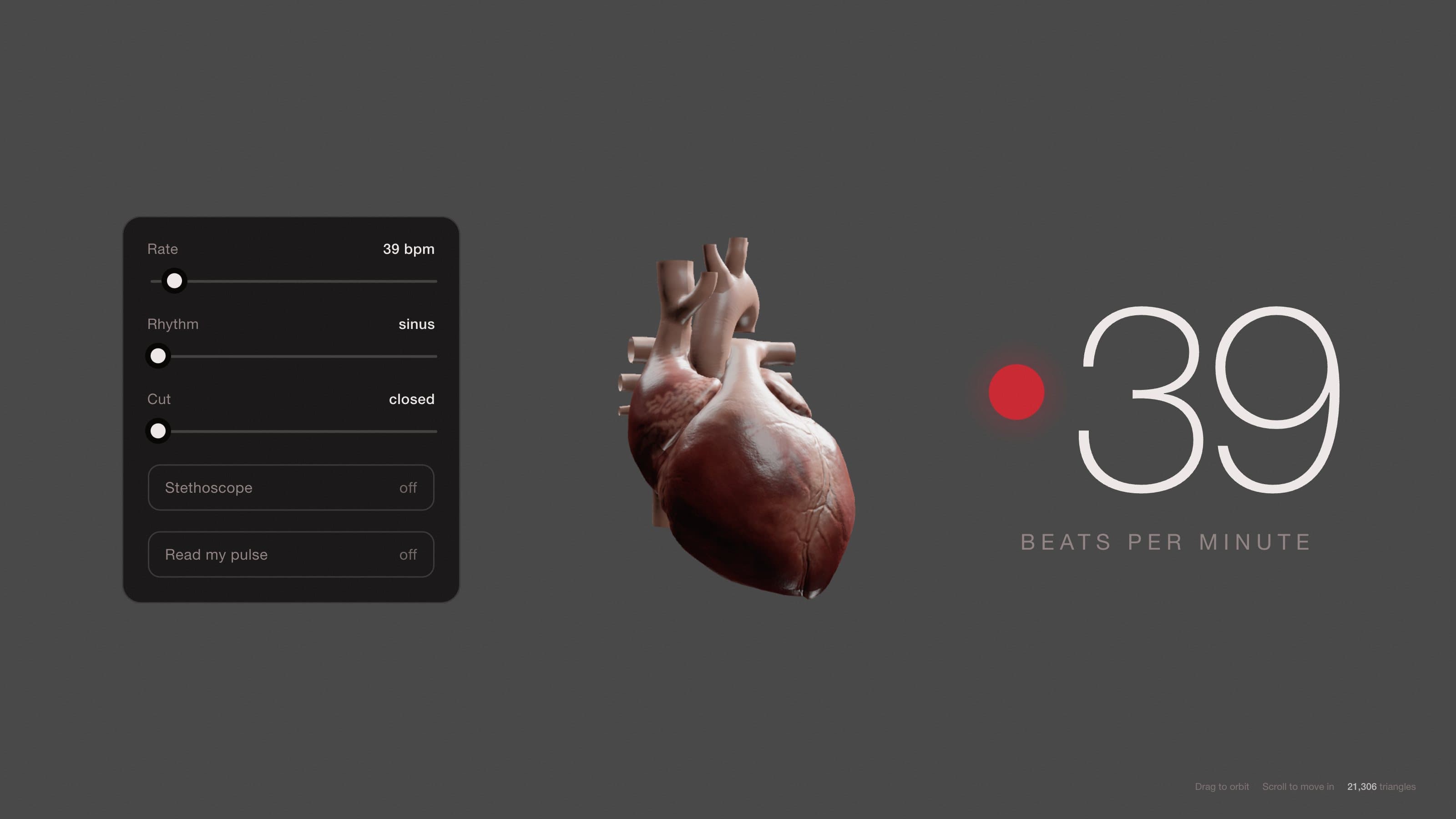
It runs in its own tab so it gets the whole screen, the sound and the full frame rate. Move and click anywhere inside it.
Overview
This interactive 3D model gives you a detailed, moving view of a human heart during a normal cardiac cycle. Watch the atria and ventricles contract and relax in real time. You control the heart rate from a slow resting beat to a faster rhythm, and you can view the heart from any angle or slice into it to see the internal chambers and valves.
The model includes structural detail (around 21,000 triangles of geometry) so you can see the wall thickness, valve positions, and septal anatomy. A live cardiac monitor displays the heart rate in beats per minute, and you can toggle on a stethoscope sound and pulse waveform to engage multiple senses while learning.
The Science
A healthy heart beats in a regular, coordinated pattern called sinus rhythm. The sinoatrial (SA) node, located in the right atrium, acts as the heart's natural pacemaker and fires electrical impulses that spread through both atria, causing them to contract first. This pushes blood into the ventricles. A brief pause allows the ventricles to fill completely before they contract forcefully to pump blood out to the body and lungs.
Heart rate varies with activity and physiology. A resting adult typically beats 60 to 100 times per minute. During exercise, stress, or illness, the rate can climb. Abnormal rhythms (arrhythmias) occur when the electrical impulses misfire or travel along irregular pathways. Understanding these patterns visually helps clinicians and patients recognize normal physiology and spot deviations early.
How to Interact
View and rotate: Click and drag on the heart to rotate it in any direction. The 3D model responds smoothly to your mouse movement so you can inspect the heart from front, back, side, or top.
Zoom in and out: Scroll with your mouse wheel to zoom into fine details like valve leaflets and coronary arteries, or zoom out for a full overview of all four chambers.
Adjust heart rate: Use the BPM slider to change the beat rate from slow (resting) to fast (exercise or stress). The counter animates in real time to show each heartbeat, and the model's contraction speed updates instantly.
Switch rhythm: Select from rhythm options to see how different electrical patterns change the contraction sequence. Sinus rhythm is the baseline normal pattern.
Cross-section view: Toggle the cut view to slice through the heart and see chambers, walls, and valve anatomy from the inside.
Sound and waveform: Turn on the stethoscope audio to hear a real cardiac sound. Enable the pulse graph to see the heart's rhythm plotted as a live waveform on a monitor-style display.
Clinical Relevance
Interactive 3D cardiac models transform how teams learn and teach heart physiology. For cardiology residencies and cardiac nursing programs, this tool lets trainees see anatomy and function together rather than relying on 2D images or diagrams. They can manipulate the model at their own pace and replay cycles until the sequence clicks.
Patient education is equally powerful. When a person learns they have a heart condition or needs a cardiac device, showing them an interactive model of their own anatomy and how treatment works builds confidence and compliance. Device companies use these models to explain pacemakers, defibrillators, and repair techniques in a way that patents and families can grasp immediately.
Med-ed publishers and hospital systems also benefit. Pre-test quizzes can embed this model so learners visualize rhythm before answering questions. The tactile, self-paced nature improves retention compared to passive video or slides.
Frequently asked questions
Can I see the inside of the heart?
Yes. Toggle the cross-section view to cut a slice through the heart model. You can then rotate and zoom to explore the chambers, septum, and valve anatomy from the inside.
What heart rates can I set?
The BPM slider lets you range from resting bradycardia (low 40s) through normal resting (60-100) and up to elevated or exercise rates (160+). The model animates the contraction speed to match your selected rate.
Is the sound realistic?
Yes. The stethoscope audio is a real recording of normal sinus rhythm. As you change the heart rate or rhythm pattern, the audio tempo adjusts to match the visual model so they stay synchronized.
Need this for your team?
Interactive cardiac models accelerate learning and boost patient confidence
Book a discovery callInteractive animations are priced per scope. Average turnaround: 15-25 days.